
המקורות לפרק
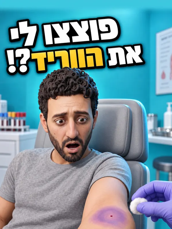
למה נשאר כתם סגול אחרי בדיקת דם?
20 טענות ו־10 מקורות. כל מה שנאמר או מוצג בפרק, לפי הסדר שבו הוא מופיע.
לצפייה בפרק ב־YouTubeבדיקת דם רגילה נלקחת בדרך כלל מווריד בקפל המרפק.
- nhs.uk
NHS 'Blood tests' (reviewed 2.11.2023): blood is taken 'usually from the inside of your elbow'. WHO 2010 p.14 (Step 3): 'inspect the antecubital fossa or forearm'.
- nhs.uk
הווריד שבאמצע קפל המרפק הוא הבחירה הראשונה: קל לדקור אותו, ובצד הפנימי עוברים מתחת לווריד עורק ועצב.
- stacks.cdc.gov
WHO 2010 p.14 (§2.2.3, Step 3): 'The median cubital vein lies between muscles and is usually the most easy to puncture. Under the basilic vein runs an artery and a nerve'. KSLM 2025 (Ann Lab Med 45:343, Fig. 4): median cubital vein 'is the first priority', 'protected by the bicipital aponeurosis'.
- stacks.cdc.gov
המחט נכנסת בזווית נמוכה, 30 מעלות או פחות, עם הצד המשופע של החוד כלפי מעלה.
- doi.org
KSLM 2025 (Needle insertion): 'at an angle of <30°, with the bevel facing upward'. WHO 2010 p.15 (Step 6): 'Enter the vein swiftly at a 30 degree angle or less'. Leicestershire NHS policy 2023: 'approximately 15-30o with the bevel uppermost'.
- doi.org
מחט רגילה לבדיקת דם עבה בערך 0.8 מילימטר.
- bd.com
WHO 2010 p.22 (Table 3.1): adult routine phlebotomy 21, 22 or 23 gauge. BD technical data sheet: '21 G thin wall' outside diameter 0.800–0.830 mm (ISO 9626:2016).
- bd.com
הווריד במרפק רחב רק כשלושה־ארבעה מילימטרים.
- doi.org
Park et al. 2024, J Vasc Access 25:1793 (abstract): ultrasound of antecubital superficial veins in 461 adults; mean vertical diameter 3.01 mm (±1.10), horizontal 4.46 mm (±1.60), depth 4.81 mm.
- doi.org
'וריד מפוצץ' הוא שם עממי. בעצם זו המטומה: דם שדלף מהווריד אל הרקמה שמסביב, מתחת לעור.
- my.clevelandclinic.org
Cleveland Clinic 'Blown Vein' (12.1.2023): 'a blood vessel that's damaged due to a needle insertion'; 'some blood leaks out and pools under your skin (hematoma)'. KSLM 2025 (Complications): 'Subcutaneous blood leakage may cause swelling or bruising at the puncture site.' Europe PMC (23.10.2026): 'blown vein' 11 records vs 1,847 for hematoma+venipuncture.
- my.clevelandclinic.org
הווריד לא מתפוצץ. המחט עושה בו חור, ולפעמים ממשיכה ויוצאת מהצד השני שלו.
- my.clevelandclinic.org
Cleveland Clinic 'Blown Vein' (Overview): the needle tip sometimes 'pokes through the other side of your vein'. WHO 2010 p.29 (Table 4.1, blood donation): cause 'Skin pierced at too great an angle – and exiting vein'. WHO p.21 (§3.1.2): a too-large needle 'will tear the vein'.
- my.clevelandclinic.org
כשהמחט נכנסת בזווית תלולה מדי, היא יכולה לצאת מהדופן האחורית של הווריד, והדם בורח החוצה.
- stacks.cdc.gov
WHO 2010 p.29 (§4.2.3, Table 4.1) haematoma causes: 'Skin pierced at too great an angle – and exiting vein'; 'Needle puncturing the vein twice during the donation'. WHO p.31 (§5, arterial): prevent haematoma 'without puncturing the far side of the vessel'. Cleveland Clinic: 'pokes through the other side of your vein'.
- stacks.cdc.gov
אם הווריד מתפצל בדיוק בנקודת הדקירה, או שהמחט גדולה מדי בשבילו, הסיכון להמטומה עולה.
- stacks.cdc.gov
WHO 2010 p.14 (Step 3): 'DO NOT insert the needle where veins are diverting, because this increases the chance of a haematoma.' WHO p.21: a needle too large 'will tear the vein and cause bleeding (haematoma)'. KSLM 2025: 'Avoid venipuncture at vein branches because of the increased risk of hematoma'.
- stacks.cdc.gov
לפעמים הווריד זז הצידה מהמחט, ולכן מותחים את העור מתחת לנקודת הדקירה כדי לקבע אותו.
- my.clevelandclinic.org
Cleveland Clinic 'Blown Vein': 'Your vein may move to the side, away from the needle, during insertion.' WHO 2010 p.15: 'Anchor the vein by holding the patient's arm and placing a thumb BELOW the venepuncture site.' KSLM 2025: 'Vein stabilization is necessary to prevent rotation during puncture' (geriatric section). Leicestershire NHS 2023: 'To immobilise the vein'.
- my.clevelandclinic.org
תזוזה קטנה של היד, ורידים שנעשים שבריריים עם הגיל, ותרופות נוגדות קרישה מגבירים את הסיכוי לכחול.
- my.clevelandclinic.org
Cleveland Clinic 'Blown Vein' (Causes): 'Movement (even slight) when the needle goes in'; 'Your veins naturally grow more fragile as you get older'. Leicestershire NHS 2023: check for anticoagulants to prevent 'haematoma through delayed haemostasis'. WHO p.54 (Table 8.1): anticoagulants under 'Extensive bleeding'.
- my.clevelandclinic.org
את הגומייה משחררים לפני שמוציאים את המחט. אחרת הלחץ בווריד דוחף דם החוצה דרך החור.
- doi.org
KSLM 2025: 'To prevent hematoma formation, release the tourniquet before needle removal'. WHO 2010 p.15: 'release the tourniquet BEFORE withdrawing the needle'. Leicestershire NHS 2023: remove bottle, then release tourniquet, 'To decrease pressure within the vein'.
- doi.org
גם בדקירה מושלמת נשאר חור בדופן הווריד, והכחול נמנע בעיקר בלחיצה עד שהדימום נעצר.
- pubmed.ncbi.nlm.nih.gov
Godwin et al. 1992, Qual Health Care 1:245 (abstract): two audits of 100 inpatients; bruising in 45, then in 25 after phlebotomists 'ensured that haemostasis had been attained before leaving the patient'. WHO 2010 p.29: cause 'Inadequate pressure after the donation'. Demirtas et al. 2026, Geriatr Nurs 69:103918 (RCT, 164 older patients on anticoagulants): 5 min pressure 'reduced the development of ecchymosis'.
- pubmed.ncbi.nlm.nih.gov
ההנחיות: ללחוץ כמה דקות עם יד ישרה, ולא לקפל את המרפק.
- stacks.cdc.gov
WHO 2010 p.15 (Step 6): 'with the arm extended and raised. Ask the patient NOT to bend the arm, because doing so causes a haematoma.' WHO p.54 (Table 8.1): 'Apply pressure to a straight arm for 3–5 minutes after drawing blood'. KSLM 2025: 'Avoid having the patient bend their arm at the elbow while applying pressure'.
- stacks.cdc.gov
סימן כחול אחרי בדיקת דם הוא דבר נפוץ: במחקר על יותר מ־4,000 בדיקות, הוא הופיע באחת מכל שמונה.
- pubmed.ncbi.nlm.nih.gov
Galena 1992, J Fam Pract 34:582 (abstract): 'Four thousand fifty venipunctures'; 'Minor bruising and hematoma were fairly common, involving 12.3% of venipunctures'.
- pubmed.ncbi.nlm.nih.gov
זה כמעט תמיד לא מסוכן, ונעלם מעצמו.
- doi.org
KSLM 2025: 'Most hematomas resolve naturally without intervention.' Cleveland Clinic 'Blown Vein': 'usually isn't dangerous or a cause for concern.' NHS 'Blood tests': bruising or swelling 'should go away after a few days'.
- doi.org
הווריד מחלים בתוך כשבועיים, ועד אז לא דוקרים בו שוב.
- my.clevelandclinic.org
Cleveland Clinic 'Blown Vein' (Outlook): 'Your blown vein will heal in about 10 to 12 days'; 'Your provider won't use that vein for IV access or blood draws until it's fully healed.' Cleveland Clinic 'Bruises': 'Most bruises fade away within two weeks'.
- my.clevelandclinic.org
הכחול משנה צבע כי הגוף מפרק את הדם שדלף: אנזים הופך את ההמוגלובין לביליוורדין ירקרק, ואחר כך לבילירובין צהוב.
- doi.org
Urakov et al. 2021, Heliyon 7:e05954 (Results): 'biliverdin, which appears greenish'; biliverdin 'is metabolized into bilirubin, which has a yellow color.' Al-Arami et al. 2025, Forensic Sci Med Pathol 21:1699 (Intro): hemoglobin 'transformed by the enzyme heme oxygenase into biliverdin', then 'converted to bilirubin by biliverdin reductase'. Langlois et al. 2015 (FSMP 11:482): heme oxygenase is 'The first step in catabolism of hemoglobin in a bruise'.
- doi.org
בהתחלה הכחול אדום־סגול, ואחר כך הוא מקבל גוונים של ירוק וצהוב, עד שהוא נעלם אחרי שבועיים־שלושה.
- doi.org
Marin et al. 2021, Sensors 21:302 (Intro, citing Langlois & Gresham 1991): 'bruises initially appear as red or pink discoloration which transforms relatively quickly into bluish/purple', 'followed by grey/brown, green, and yellow hues, until the resolution 2–3 weeks post injury'. Bilirubin 'typically peaks at 3–5 days'. Langlois & Gresham 1991 (FSI 50:227): 'a bruise with a yellow colour was more than 18 h old'.
- doi.org
הדם שדלף לא כחול. הוא אדום כהה, והגוון הכחלחל נוצר מהאור שעובר דרך העור.
- doi.org
Urakov et al. 2021, Heliyon (Introduction): 'deoxyhemoglobin is dark red'; 'The blue appearance of bruises is due to the higher reflection of the blue light component'.
- doi.org
מצאתם טעות? כתבו לנו ל־hello@beetzem.com.